A blurred spot can still reveal a very precise centre
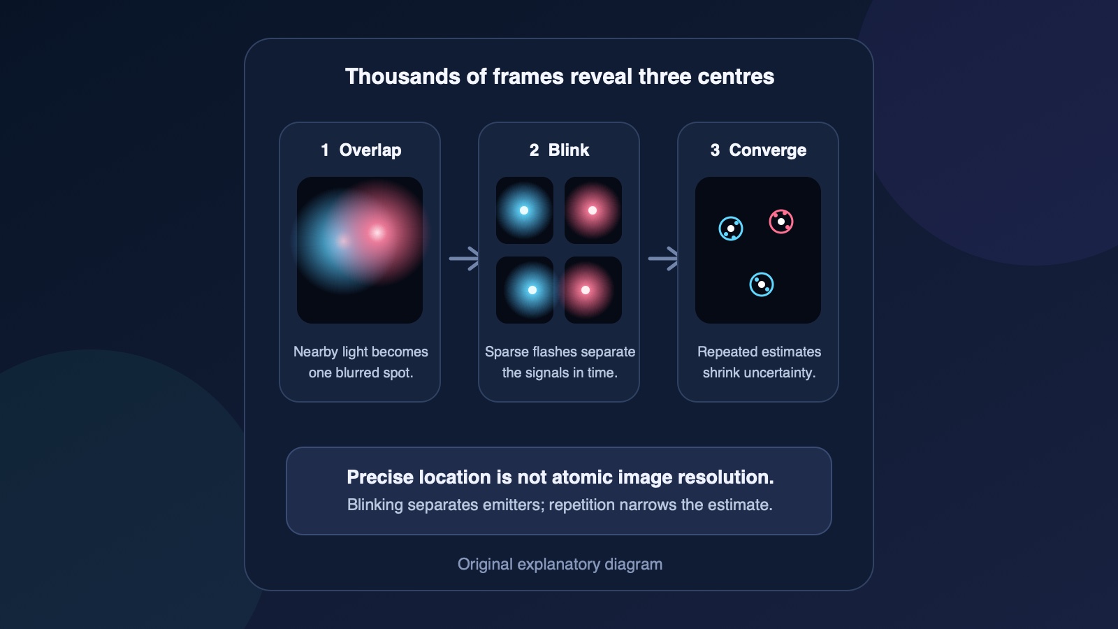
Visible light spreads as it passes through a microscope, so two emitters closer than roughly 200 to 300 nanometres can merge into one blurred patch. That diffraction limit restricts the detail a conventional light microscope can separate. It does not prevent a computer from estimating the centre of one isolated patch much more precisely.
Think of many darts landing around a hidden bullseye. One dart gives a rough clue; thousands can reveal the centre of their cluster with much smaller statistical uncertainty. Single-molecule localization microscopy uses that principle with photons, provided nearby light emitters can be made to take turns instead of glowing together.
Blinking separates crowded signals in time
STORM, short for stochastic optical reconstruction microscopy, records a long sequence of frames in which only a sparse set of fluorescent labels is switched on. Because their blurred spots no longer overlap, software can estimate each centre. Combining many localized points reconstructs detail that the original crowded view could not separate.
Conventional fluorescent dyes create practical limits. They can bleach, meaning their light output is permanently damaged, and multicolour work may need different lasers, chemical buffers or sequential imaging rounds. A probe that turns itself on and off for a very long time could supply far more position estimates with a simpler optical setup.
U-STORM engineers the blinking into a tiny crystal
The new probes are upconverting nanoparticles roughly 10 nanometres across. Ytterbium ions absorb near-infrared light and pass energy through the particle to thulium or erbium ions that emit visible blue or red light. By tuning those ingredients and adding a thin shell, the researchers made the particles switch spontaneously between bright and dark states.
The paper reports duty cycles as low as about 0.9%, meaning a particle stays dark most of the time and briefly appears as a separable signal. Under continuous near-infrared excitation, tested particles kept blinking for ten hours without the observed photobleaching or statistical ageing that would progressively remove useful flashes.
Eighty-eight thousand estimates shrink one uncertainty
A single bright event still located the nanoparticle only to the ordinary tens-of-nanometres range. The gain came from repeatedly measuring the same emitter. With about 88,000 localizations, the uncertainty in the mean position fell with the square root of the number of observations, reaching a reported 0.62 ångström. One ångström is one ten-billionth of a metre.
That number is smaller than the width of many atoms, but it describes localization precision: how tightly repeated estimates cluster around a centre. Image resolution asks whether two neighbouring structures can be distinguished. Accuracy asks whether the estimated centre is also free from drift, calibration errors and other bias. A very precise coordinate is not automatically an atomic photograph.
The experiment resolved nanoparticles and protein arrangements, not atoms
The researchers checked the optical estimates against scanning electron microscope images of the same field. U-STORM separated eight nanoparticles hidden inside one diffraction-limited spot and measured centre-to-centre separations down to about 14 nanometres. That validation shows useful agreement between the reconstructed coordinates and physical particle positions.
The team also used blue- and red-emitting probes in one imaging round to map epidermal growth factor receptor dimers and multimers on cell membranes. Those results support single-protein-scale biological imaging with a simpler excitation arrangement. They do not show the internal atoms of a protein, and the 10-nanometre labels themselves remain part of what a biological experiment can resolve.
The next milestone is reliable biology, not a smaller headline number
For U-STORM to become a widely useful tool, independent laboratories need to reproduce the blinking, precision and multicolour results. The particles must become brighter or smaller where needed, attach selectively without disturbing their targets, work across thicker and living samples, and preserve accuracy when the specimen moves or the microscope drifts.
A result that fails those replication, labelling or live-sample tests would delay the promise even if one calibration particle still produces an impressive uncertainty value. Success would look different: routine maps of protein organisation that competing methods cannot obtain as simply. The real frontier is not seeing atoms through ordinary glass. It is learning how repeated, durable flashes can turn blurred light into trustworthy molecular coordinates.
Sources and further reading
- Nature Nanotechnology: open-access U-STORM paper and experimental results ↗
- MIT and Broad Institute: institutional research summary and visual explanation ↗
- Journal of Cell Science review: resolution, localization precision and super-resolution limits ↗
- Journal of Microscopy review: localization is distinct from image resolution ↗
This article was written for Curiosity Desk. We do not copy other publishers or invent quotes. If a material error is found, we correct it openly.
Read the full standards →One answer should lead to a better question
Bring your curiosity to the group
Curious Minds is our public Facebook community for surprising science, strange history, Australian wildlife and everyday questions. No copied posts, no personal-friend invitations and no link dumping.
- Three self-contained discussion prompts each week
- Sourced answers and honest uncertainty
- Respectful conversation without spam